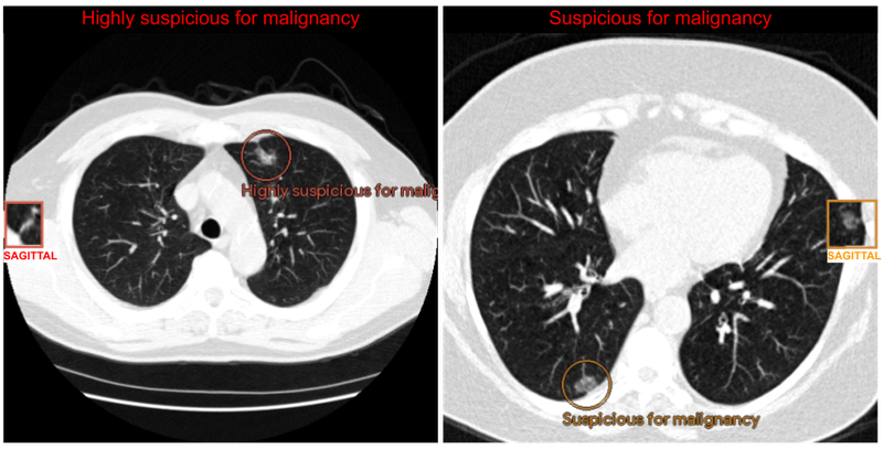
Introducing a generalizable user-centric interface to assist radiologists leverage machine studying fashions for lung most cancers screening. The system takes computed tomography (CT) imaging as enter and outputs a most cancers suspicion score together with the corresponding areas of curiosity.
Lung most cancers is the main explanation for cancer-related deaths globally with 1.8 million deaths reported in 2020. Late prognosis dramatically reduces the possibilities of survival. Lung most cancers screening by way of computed tomography (CT), which gives an in depth 3D picture of the lungs, has been proven to cut back mortality in high-risk populations by a minimum of 20% by detecting potential indicators of cancers earlier. Within the US, screening entails annual scans, with some nations or instances recommending kind of frequent scans.
The United States Preventive Providers Activity Pressure just lately expanded lung most cancers screening suggestions by roughly 80%, which is anticipated to extend screening entry for girls and racial and ethnic minority teams. Nevertheless, false positives (i.e., incorrectly reporting a possible most cancers in a cancer-free affected person) may cause nervousness and result in pointless procedures for sufferers whereas growing prices for the healthcare system. Furthermore, effectivity in screening a lot of people could be difficult relying on healthcare infrastructure and radiologist availability.
At Google we’ve beforehand developed machine studying (ML) fashions for lung most cancers detection, and have evaluated their skill to routinely detect and classify areas that present indicators of potential most cancers. Efficiency has been proven to be corresponding to that of specialists in detecting doable most cancers. Whereas they’ve achieved excessive efficiency, successfully speaking findings in sensible environments is important to understand their full potential.
To that finish, in “Assistive AI in Lung Most cancers Screening: A Retrospective Multinational Examine within the US and Japan”, printed in Radiology AI, we examine how ML fashions can successfully talk findings to radiologists. We additionally introduce a generalizable user-centric interface to assist radiologists leverage such fashions for lung most cancers screening. The system takes CT imaging as enter and outputs a most cancers suspicion score utilizing 4 classes (no suspicion, in all probability benign, suspicious, extremely suspicious) together with the corresponding areas of curiosity. We consider the system’s utility in enhancing clinician efficiency by means of randomized reader research in each the US and Japan, utilizing the native most cancers scoring techniques (Lung-RADSs V1.1 and Sendai Rating) and picture viewers that mimic sensible settings. We discovered that reader specificity will increase with mannequin help in each reader research. To speed up progress in conducting comparable research with ML fashions, we’ve open-sourced code to course of CT pictures and generate pictures suitable with the image archiving and communication system (PACS) utilized by radiologists.
Growing an interface to speak mannequin outcomes
Integrating ML fashions into radiologist workflows entails understanding the nuances and objectives of their duties to meaningfully help them. Within the case of lung most cancers screening, hospitals observe numerous country-specific pointers which can be commonly up to date. For instance, within the US, Lung-RADs V1.1 assigns an alpha-numeric rating to point the lung most cancers threat and follow-up suggestions. When assessing sufferers, radiologists load the CT of their workstation to learn the case, discover lung nodules or lesions, and apply set pointers to find out follow-up selections.
Our first step was to enhance the beforehand developed ML fashions by means of extra coaching information and architectural enhancements, together with self-attention. Then, as an alternative of concentrating on particular pointers, we experimented with a complementary approach of speaking AI outcomes unbiased of pointers or their specific variations. Particularly, the system output gives a suspicion score and localization (areas of curiosity) for the person to think about together with their very own particular pointers. The interface produces output pictures instantly related to the CT research, requiring no modifications to the person’s workstation. The radiologist solely must evaluate a small set of extra pictures. There isn’t a different change to their system or interplay with the system.

Instance of the assistive lung most cancers screening system outputs. Outcomes for the radiologist’s analysis are visualized on the situation of the CT quantity the place the suspicious lesion is discovered. The general suspicion is displayed on the high of the CT pictures. Circles spotlight the suspicious lesions whereas squares present a rendering of the identical lesion from a unique perspective, referred to as a sagittal view.
The assistive lung most cancers screening system includes 13 fashions and has a high-level structure much like the end-to-end system utilized in prior work. The fashions coordinate with one another to first section the lungs, get hold of an general evaluation, find three suspicious areas, then use the data to assign a suspicion score to every area. The system was deployed on Google Cloud utilizing a Google Kubernetes Engine (GKE) that pulled the pictures, ran the ML fashions, and supplied outcomes. This enables scalability and instantly connects to servers the place the pictures are saved in DICOM shops.

Define of the Google Cloud deployment of the assistive lung most cancers screening system and the directional calling stream for the person elements that serve the pictures and compute outcomes. Pictures are served to the viewer and to the system utilizing Google Cloud companies. The system is run on a Google Kubernetes Engine that pulls the pictures, processes them, and writes them again into the DICOM retailer.
Reader research
To guage the system’s utility in enhancing scientific efficiency, we performed two reader research (i.e., experiments designed to evaluate scientific efficiency evaluating professional efficiency with and with out the help of a know-how) with 12 radiologists utilizing pre-existing, de-identified CT scans. We introduced 627 difficult instances to six US-based and 6 Japan-based radiologists. Within the experimental setup, readers have been divided into two teams that learn every case twice, with and with out help from the mannequin. Readers have been requested to use scoring pointers they sometimes use of their scientific observe and report their general suspicion of most cancers for every case. We then in contrast the outcomes of the reader’s responses to measure the influence of the mannequin on their workflow and selections. The rating and suspicion stage have been judged towards the precise most cancers outcomes of the people to measure sensitivity, specificity, and space underneath the ROC curve (AUC) values. These have been in contrast with and with out help.

A multi-case multi-reader research entails every case being reviewed by every reader twice, as soon as with ML system help and as soon as with out. On this visualization one reader first opinions Set A with out help (blue) after which with help (orange) after a wash-out interval. A second reader group follows the other path by studying the identical set of instances Set A with help first. Readers are randomized to those teams to take away the impact of ordering.
The power to conduct these research utilizing the identical interface highlights its generalizability to fully completely different most cancers scoring techniques, and the generalization of the mannequin and assistive functionality to completely different affected person populations. Our research outcomes demonstrated that when radiologists used the system of their scientific analysis, they’d an elevated skill to accurately determine lung pictures with out actionable lung most cancers findings (i.e., specificity) by an absolute 5–7% in comparison with once they didn’t use the assistive system. This doubtlessly implies that for each 15–20 sufferers screened, one might be able to keep away from pointless follow-up procedures, thus lowering their nervousness and the burden on the well being care system. This will, in flip, assist enhance the sustainability of lung most cancers screening applications, notably as extra individuals change into eligible for screening.

Reader specificity will increase with ML mannequin help in each the US-based and Japan-based reader research. Specificity values have been derived from reader scores from actionable findings (one thing suspicious was discovered) versus no actionable findings, in contrast towards the true most cancers final result of the person. Underneath mannequin help, readers flagged fewer cancer-negative people for follow-up visits. Sensitivity for most cancers constructive people remained the identical.
Translating this into real-world influence by means of partnership
The system outcomes show the potential for fewer follow-up visits, diminished nervousness, as effectively decrease general prices for lung most cancers screening. In an effort to translate this analysis into real-world scientific influence, we’re working with: DeepHealth, a number one AI-powered well being informatics supplier; and Apollo Radiology Worldwide a number one supplier of Radiology companies in India to discover paths for incorporating this method into future merchandise. As well as, we want to assist different researchers finding out how greatest to combine ML mannequin outcomes into scientific workflows by open sourcing code used for the reader research and incorporating the insights described on this weblog. We hope that this can assist speed up medical imaging researchers trying to conduct reader research for his or her AI fashions, and catalyze translational analysis within the subject.
Acknowledgements
Key contributors to this challenge embrace Corbin Cunningham, Zaid Nabulsi, Ryan Najafi, Jie Yang, Charles Lau, Joseph R. Ledsam, Wenxing Ye, Diego Ardila, Scott M. McKinney, Rory Pilgrim, Hiroaki Saito, Yasuteru Shimamura, Mozziyar Etemadi, Yun Liu, David Melnick, Sunny Jansen, Nadia Harhen, David P. Nadich, Mikhail Fomitchev, Ziyad Helali, Shabir Adeel, Greg S. Corrado, Lily Peng, Daniel Tse, Shravya Shetty, Shruthi Prabhakara, Neeral Beladia, and Krish Eswaran. Because of Arnav Agharwal and Andrew Sellergren for his or her open sourcing help and Vivek Natarajan and Michael D. Howell for his or her suggestions. Honest appreciation additionally goes to the radiologists who enabled this work with their picture interpretation and annotation efforts all through the research, and Jonny Wong and Carli Sampson for coordinating the reader research.
Introducing a generalizable user-centric interface to assist radiologists leverage machine studying fashions for lung most cancers screening. The system takes computed tomography (CT) imaging as enter and outputs a most cancers suspicion score together with the corresponding areas of curiosity.
Lung most cancers is the main explanation for cancer-related deaths globally with 1.8 million deaths reported in 2020. Late prognosis dramatically reduces the possibilities of survival. Lung most cancers screening by way of computed tomography (CT), which gives an in depth 3D picture of the lungs, has been proven to cut back mortality in high-risk populations by a minimum of 20% by detecting potential indicators of cancers earlier. Within the US, screening entails annual scans, with some nations or instances recommending kind of frequent scans.
The United States Preventive Providers Activity Pressure just lately expanded lung most cancers screening suggestions by roughly 80%, which is anticipated to extend screening entry for girls and racial and ethnic minority teams. Nevertheless, false positives (i.e., incorrectly reporting a possible most cancers in a cancer-free affected person) may cause nervousness and result in pointless procedures for sufferers whereas growing prices for the healthcare system. Furthermore, effectivity in screening a lot of people could be difficult relying on healthcare infrastructure and radiologist availability.
At Google we’ve beforehand developed machine studying (ML) fashions for lung most cancers detection, and have evaluated their skill to routinely detect and classify areas that present indicators of potential most cancers. Efficiency has been proven to be corresponding to that of specialists in detecting doable most cancers. Whereas they’ve achieved excessive efficiency, successfully speaking findings in sensible environments is important to understand their full potential.
To that finish, in “Assistive AI in Lung Most cancers Screening: A Retrospective Multinational Examine within the US and Japan”, printed in Radiology AI, we examine how ML fashions can successfully talk findings to radiologists. We additionally introduce a generalizable user-centric interface to assist radiologists leverage such fashions for lung most cancers screening. The system takes CT imaging as enter and outputs a most cancers suspicion score utilizing 4 classes (no suspicion, in all probability benign, suspicious, extremely suspicious) together with the corresponding areas of curiosity. We consider the system’s utility in enhancing clinician efficiency by means of randomized reader research in each the US and Japan, utilizing the native most cancers scoring techniques (Lung-RADSs V1.1 and Sendai Rating) and picture viewers that mimic sensible settings. We discovered that reader specificity will increase with mannequin help in each reader research. To speed up progress in conducting comparable research with ML fashions, we’ve open-sourced code to course of CT pictures and generate pictures suitable with the image archiving and communication system (PACS) utilized by radiologists.
Growing an interface to speak mannequin outcomes
Integrating ML fashions into radiologist workflows entails understanding the nuances and objectives of their duties to meaningfully help them. Within the case of lung most cancers screening, hospitals observe numerous country-specific pointers which can be commonly up to date. For instance, within the US, Lung-RADs V1.1 assigns an alpha-numeric rating to point the lung most cancers threat and follow-up suggestions. When assessing sufferers, radiologists load the CT of their workstation to learn the case, discover lung nodules or lesions, and apply set pointers to find out follow-up selections.
Our first step was to enhance the beforehand developed ML fashions by means of extra coaching information and architectural enhancements, together with self-attention. Then, as an alternative of concentrating on particular pointers, we experimented with a complementary approach of speaking AI outcomes unbiased of pointers or their specific variations. Particularly, the system output gives a suspicion score and localization (areas of curiosity) for the person to think about together with their very own particular pointers. The interface produces output pictures instantly related to the CT research, requiring no modifications to the person’s workstation. The radiologist solely must evaluate a small set of extra pictures. There isn’t a different change to their system or interplay with the system.

Instance of the assistive lung most cancers screening system outputs. Outcomes for the radiologist’s analysis are visualized on the situation of the CT quantity the place the suspicious lesion is discovered. The general suspicion is displayed on the high of the CT pictures. Circles spotlight the suspicious lesions whereas squares present a rendering of the identical lesion from a unique perspective, referred to as a sagittal view.
The assistive lung most cancers screening system includes 13 fashions and has a high-level structure much like the end-to-end system utilized in prior work. The fashions coordinate with one another to first section the lungs, get hold of an general evaluation, find three suspicious areas, then use the data to assign a suspicion score to every area. The system was deployed on Google Cloud utilizing a Google Kubernetes Engine (GKE) that pulled the pictures, ran the ML fashions, and supplied outcomes. This enables scalability and instantly connects to servers the place the pictures are saved in DICOM shops.

Define of the Google Cloud deployment of the assistive lung most cancers screening system and the directional calling stream for the person elements that serve the pictures and compute outcomes. Pictures are served to the viewer and to the system utilizing Google Cloud companies. The system is run on a Google Kubernetes Engine that pulls the pictures, processes them, and writes them again into the DICOM retailer.
Reader research
To guage the system’s utility in enhancing scientific efficiency, we performed two reader research (i.e., experiments designed to evaluate scientific efficiency evaluating professional efficiency with and with out the help of a know-how) with 12 radiologists utilizing pre-existing, de-identified CT scans. We introduced 627 difficult instances to six US-based and 6 Japan-based radiologists. Within the experimental setup, readers have been divided into two teams that learn every case twice, with and with out help from the mannequin. Readers have been requested to use scoring pointers they sometimes use of their scientific observe and report their general suspicion of most cancers for every case. We then in contrast the outcomes of the reader’s responses to measure the influence of the mannequin on their workflow and selections. The rating and suspicion stage have been judged towards the precise most cancers outcomes of the people to measure sensitivity, specificity, and space underneath the ROC curve (AUC) values. These have been in contrast with and with out help.

A multi-case multi-reader research entails every case being reviewed by every reader twice, as soon as with ML system help and as soon as with out. On this visualization one reader first opinions Set A with out help (blue) after which with help (orange) after a wash-out interval. A second reader group follows the other path by studying the identical set of instances Set A with help first. Readers are randomized to those teams to take away the impact of ordering.
The power to conduct these research utilizing the identical interface highlights its generalizability to fully completely different most cancers scoring techniques, and the generalization of the mannequin and assistive functionality to completely different affected person populations. Our research outcomes demonstrated that when radiologists used the system of their scientific analysis, they’d an elevated skill to accurately determine lung pictures with out actionable lung most cancers findings (i.e., specificity) by an absolute 5–7% in comparison with once they didn’t use the assistive system. This doubtlessly implies that for each 15–20 sufferers screened, one might be able to keep away from pointless follow-up procedures, thus lowering their nervousness and the burden on the well being care system. This will, in flip, assist enhance the sustainability of lung most cancers screening applications, notably as extra individuals change into eligible for screening.

Reader specificity will increase with ML mannequin help in each the US-based and Japan-based reader research. Specificity values have been derived from reader scores from actionable findings (one thing suspicious was discovered) versus no actionable findings, in contrast towards the true most cancers final result of the person. Underneath mannequin help, readers flagged fewer cancer-negative people for follow-up visits. Sensitivity for most cancers constructive people remained the identical.
Translating this into real-world influence by means of partnership
The system outcomes show the potential for fewer follow-up visits, diminished nervousness, as effectively decrease general prices for lung most cancers screening. In an effort to translate this analysis into real-world scientific influence, we’re working with: DeepHealth, a number one AI-powered well being informatics supplier; and Apollo Radiology Worldwide a number one supplier of Radiology companies in India to discover paths for incorporating this method into future merchandise. As well as, we want to assist different researchers finding out how greatest to combine ML mannequin outcomes into scientific workflows by open sourcing code used for the reader research and incorporating the insights described on this weblog. We hope that this can assist speed up medical imaging researchers trying to conduct reader research for his or her AI fashions, and catalyze translational analysis within the subject.
Acknowledgements
Key contributors to this challenge embrace Corbin Cunningham, Zaid Nabulsi, Ryan Najafi, Jie Yang, Charles Lau, Joseph R. Ledsam, Wenxing Ye, Diego Ardila, Scott M. McKinney, Rory Pilgrim, Hiroaki Saito, Yasuteru Shimamura, Mozziyar Etemadi, Yun Liu, David Melnick, Sunny Jansen, Nadia Harhen, David P. Nadich, Mikhail Fomitchev, Ziyad Helali, Shabir Adeel, Greg S. Corrado, Lily Peng, Daniel Tse, Shravya Shetty, Shruthi Prabhakara, Neeral Beladia, and Krish Eswaran. Because of Arnav Agharwal and Andrew Sellergren for his or her open sourcing help and Vivek Natarajan and Michael D. Howell for his or her suggestions. Honest appreciation additionally goes to the radiologists who enabled this work with their picture interpretation and annotation efforts all through the research, and Jonny Wong and Carli Sampson for coordinating the reader research.


