


Google analysis releases two new research-use-only instruments for coaching medical imaging fashions for dermatology and pathology, constructing on its domain-specific embeddings.
There’s a worldwide scarcity of entry to medical imaging skilled interpretation throughout specialties together with radiology, dermatology and pathology. Machine studying (ML) know-how may help ease this burden by powering instruments that allow medical doctors to interpret these pictures extra precisely and effectively. Nonetheless, the event and implementation of such ML instruments are sometimes restricted by the supply of high-quality information, ML experience, and computational assets.
One option to catalyze using ML for medical imaging is by way of domain-specific fashions that make the most of deep studying (DL) to seize the knowledge in medical pictures as compressed numerical vectors (referred to as embeddings). These embeddings symbolize a kind of pre-learned understanding of the vital options in a picture. Figuring out patterns within the embeddings reduces the quantity of information, experience, and compute wanted to coach performant fashions as in comparison with working with high-dimensional information, resembling pictures, straight. Certainly, these embeddings can be utilized to carry out quite a lot of downstream duties inside the specialised area (see animated graphic beneath). This framework of leveraging pre-learned understanding to unravel associated duties is much like that of a seasoned guitar participant rapidly studying a brand new music by ear. As a result of the guitar participant has already constructed up a basis of ability and understanding, they will rapidly choose up the patterns and groove of a brand new music.
Path Basis is used to transform a small dataset of (picture, label) pairs into (embedding, label) pairs. These pairs can then be used to coach a task-specific classifier utilizing a linear probe, (i.e., a light-weight linear classifier) as represented on this graphic, or different kinds of fashions utilizing the embeddings as enter.
As soon as the linear probe is skilled, it may be used to make predictions on embeddings from new pictures. These predictions might be in comparison with floor reality info with a view to consider the linear probe’s efficiency.
In an effort to make such a embedding mannequin obtainable and drive additional improvement of ML instruments in medical imaging, we’re excited to launch two domain-specific instruments for analysis use: Derm Basis and Path Basis. This follows on the robust response we’ve already obtained from researchers utilizing the CXR Basis embedding device for chest radiographs and represents a portion of our increasing analysis choices throughout a number of medical-specialized modalities. These embedding instruments take a picture as enter and produce a numerical vector (the embedding) that’s specialised to the domains of dermatology and digital pathology pictures, respectively. By operating a dataset of chest X-ray, dermatology, or pathology pictures by means of the respective embedding device, researchers can acquire embeddings for their very own pictures, and use these embeddings to rapidly develop new fashions for his or her functions.
Path Basis
In “Area-specific optimization and numerous analysis of self-supervised fashions for histopathology”, we confirmed that self-supervised studying (SSL) fashions for pathology pictures outperform conventional pre-training approaches and allow environment friendly coaching of classifiers for downstream duties. This effort centered on hematoxylin and eosin (H&E) stained slides, the principal tissue stain in diagnostic pathology that allows pathologists to visualise mobile options underneath a microscope. The efficiency of linear classifiers skilled utilizing the output of the SSL fashions matched that of prior DL fashions skilled on orders of magnitude extra labeled information.
On account of substantial variations between digital pathology pictures and “pure picture” photographs, this work concerned a number of pathology-specific optimizations throughout mannequin coaching. One key ingredient is that whole-slide pictures (WSIs) in pathology might be 100,000 pixels throughout (1000’s of occasions bigger than typical smartphone photographs) and are analyzed by consultants at a number of magnifications (zoom ranges). As such, the WSIs are usually damaged down into smaller tiles or patches for pc imaginative and prescient and DL functions. The ensuing pictures are info dense with cells or tissue constructions distributed all through the body as a substitute of getting distinct semantic objects or foreground vs. background variations, thus creating distinctive challenges for strong SSL and have extraction. Moreover, bodily (e.g., slicing) and chemical (e.g., fixing and staining) processes used to arrange the samples can affect picture look dramatically.
Taking these vital features into consideration, pathology-specific SSL optimizations included serving to the mannequin study stain-agnostic options, generalizing the mannequin to patches from a number of magnifications, augmenting the information to imitate scanning and picture submit processing, and customized information balancing to enhance enter heterogeneity for SSL coaching. These approaches have been extensively evaluated utilizing a broad set of benchmark duties involving 17 completely different tissue varieties over 12 completely different duties.
Using the imaginative and prescient transformer (ViT-S/16) structure, Path Basis was chosen as one of the best performing mannequin from the optimization and analysis course of described above (and illustrated within the determine beneath). This mannequin thus offers an vital steadiness between efficiency and mannequin measurement to allow precious and scalable use in producing embeddings over the various particular person picture patches of huge pathology WSIs.
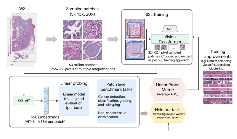
SSL coaching with pathology-specific optimizations for Path Basis.
The worth of domain-specific picture representations can be seen within the determine beneath, which reveals the linear probing efficiency enchancment of Path Basis (as measured by AUROC) in comparison with conventional pre-training on pure pictures (ImageNet-21k). This consists of analysis for duties resembling metastatic breast most cancers detection in lymph nodes, prostate most cancers grading, and breast most cancers grading, amongst others.

Path Basis embeddings considerably outperform conventional ImageNet embeddings as evaluated by linear probing throughout a number of analysis duties in histopathology.
Derm Basis
Derm Basis is an embedding device derived from our analysis in making use of DL to interpret pictures of dermatology circumstances and consists of our latest work that provides enhancements to generalize higher to new datasets. On account of its dermatology-specific pre-training it has a latent understanding of options current in pictures of pores and skin circumstances and can be utilized to rapidly develop fashions to categorise pores and skin circumstances. The mannequin underlying the API is a BiT ResNet-101×3 skilled in two phases. The primary pre-training stage makes use of contrastive studying, much like ConVIRT, to coach on numerous image-text pairs from the web. Within the second stage, the picture part of this pre-trained mannequin is then fine-tuned for situation classification utilizing medical datasets, resembling these from teledermatology providers.
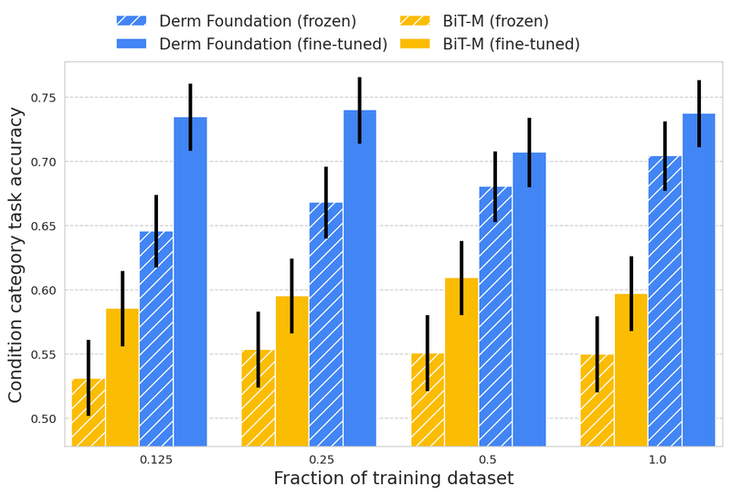
In contrast to histopathology pictures, dermatology pictures extra carefully resemble the real-world pictures used to coach a lot of in the present day’s pc imaginative and prescient fashions. Nonetheless, for specialised dermatology duties, making a high-quality mannequin should require a big dataset. With Derm Basis, researchers can use their very own smaller dataset to retrieve domain-specific embeddings, and use these to construct smaller fashions (e.g., linear classifiers or different small non-linear fashions) that allow them to validate their analysis or product concepts. To judge this method, we skilled fashions on a downstream job utilizing teledermatology information. Mannequin coaching concerned various dataset sizes (12.5%, 25%, 50%, 100%) to check embedding-based linear classifiers in opposition to fine-tuning.
The modeling variants thought-about have been:
- A linear classifier on frozen embeddings from BiT-M (an ordinary pre-trained picture mannequin)
- Wonderful-tuned model of BiT-M with an additional dense layer for the downstream job
- A linear classifier on frozen embeddings from the Derm Basis API
- Wonderful-tuned model of the mannequin underlying the Derm Basis API with an additional layer for the downstream job
We discovered that fashions constructed on high of the Derm Basis embeddings for dermatology-related duties achieved considerably larger high quality than these constructed solely on embeddings or nice tuned from BiT-M. This benefit was discovered to be most pronounced for smaller coaching dataset sizes.
These outcomes exhibit that the Derm Basis tooI can function a helpful place to begin to speed up skin-related modeling duties. We purpose to allow different researchers to construct on the underlying options and representations of dermatology that the mannequin has discovered.
These outcomes exhibit that the Derm Basis tooI can function a helpful place to begin to speed up skin-related modeling duties. We purpose to allow different researchers to construct on the underlying options and representations of dermatology that the mannequin has discovered.
Nonetheless, there are limitations with this evaluation. We’re nonetheless exploring how properly these embeddings generalize throughout job varieties, affected person populations, and picture settings. Downstream fashions constructed utilizing Derm Basis nonetheless require cautious analysis to know their anticipated efficiency within the supposed setting.
Entry Path and Derm Basis
We envision that the Derm Basis and Path Basis embedding instruments will allow a spread of use circumstances, together with environment friendly improvement of fashions for diagnostic duties, high quality assurance and pre-analytical workflow enhancements, picture indexing and curation, and biomarker discovery and validation. We’re releasing each instruments to the analysis group to allow them to discover the utility of the embeddings for their very own dermatology and pathology information.
To get entry, please signal as much as every device’s phrases of service utilizing the next Google Types.
After having access to every device, you should use the API to retrieve embeddings from dermatology pictures or digital pathology pictures saved in Google Cloud. Accepted customers who’re simply curious to see the mannequin and embeddings in motion can use the offered instance Colab notebooks to coach fashions utilizing public information for classifying six frequent pores and skin circumstances or figuring out tumors in histopathology patches. We look ahead to seeing the vary of use-cases these instruments can unlock.
Acknowledgements
We wish to thank the various collaborators who helped make this work doable together with Yun Liu, Can Kirmizi, Fereshteh Mahvar, Bram Sterling, Arman Tajback, Kenneth Philbrik, Arnav Agharwal, Aurora Cheung, Andrew Sellergren, Boris Babenko, Basil Mustafa, Jan Freyberg, Terry Spitz, Yuan Liu, Pinal Bavishi, Ayush Jain, Amit Talreja, Rajeev Rikhye, Abbi Ward, Jeremy Lai, Faruk Ahmed, Supriya Vijay,Tiam Jaroensri, Jessica Bathroom, Saurabh Vyawahare, Saloni Agarwal, Ellery Wulczyn, Jonathan Krause, Fayaz Jamil, Tom Small, Annisah Um’rani, Lauren Winer, Sami Lachgar, Yossi Matias, Greg Corrado, and Dale Webster.
Google analysis releases two new research-use-only instruments for coaching medical imaging fashions for dermatology and pathology, constructing on its domain-specific embeddings.
There’s a worldwide scarcity of entry to medical imaging skilled interpretation throughout specialties together with radiology, dermatology and pathology. Machine studying (ML) know-how may help ease this burden by powering instruments that allow medical doctors to interpret these pictures extra precisely and effectively. Nonetheless, the event and implementation of such ML instruments are sometimes restricted by the supply of high-quality information, ML experience, and computational assets.
One option to catalyze using ML for medical imaging is by way of domain-specific fashions that make the most of deep studying (DL) to seize the knowledge in medical pictures as compressed numerical vectors (referred to as embeddings). These embeddings symbolize a kind of pre-learned understanding of the vital options in a picture. Figuring out patterns within the embeddings reduces the quantity of information, experience, and compute wanted to coach performant fashions as in comparison with working with high-dimensional information, resembling pictures, straight. Certainly, these embeddings can be utilized to carry out quite a lot of downstream duties inside the specialised area (see animated graphic beneath). This framework of leveraging pre-learned understanding to unravel associated duties is much like that of a seasoned guitar participant rapidly studying a brand new music by ear. As a result of the guitar participant has already constructed up a basis of ability and understanding, they will rapidly choose up the patterns and groove of a brand new music.
Path Basis is used to transform a small dataset of (picture, label) pairs into (embedding, label) pairs. These pairs can then be used to coach a task-specific classifier utilizing a linear probe, (i.e., a light-weight linear classifier) as represented on this graphic, or different kinds of fashions utilizing the embeddings as enter.
As soon as the linear probe is skilled, it may be used to make predictions on embeddings from new pictures. These predictions might be in comparison with floor reality info with a view to consider the linear probe’s efficiency.
In an effort to make such a embedding mannequin obtainable and drive additional improvement of ML instruments in medical imaging, we’re excited to launch two domain-specific instruments for analysis use: Derm Basis and Path Basis. This follows on the robust response we’ve already obtained from researchers utilizing the CXR Basis embedding device for chest radiographs and represents a portion of our increasing analysis choices throughout a number of medical-specialized modalities. These embedding instruments take a picture as enter and produce a numerical vector (the embedding) that’s specialised to the domains of dermatology and digital pathology pictures, respectively. By operating a dataset of chest X-ray, dermatology, or pathology pictures by means of the respective embedding device, researchers can acquire embeddings for their very own pictures, and use these embeddings to rapidly develop new fashions for his or her functions.
Path Basis
In “Area-specific optimization and numerous analysis of self-supervised fashions for histopathology”, we confirmed that self-supervised studying (SSL) fashions for pathology pictures outperform conventional pre-training approaches and allow environment friendly coaching of classifiers for downstream duties. This effort centered on hematoxylin and eosin (H&E) stained slides, the principal tissue stain in diagnostic pathology that allows pathologists to visualise mobile options underneath a microscope. The efficiency of linear classifiers skilled utilizing the output of the SSL fashions matched that of prior DL fashions skilled on orders of magnitude extra labeled information.
On account of substantial variations between digital pathology pictures and “pure picture” photographs, this work concerned a number of pathology-specific optimizations throughout mannequin coaching. One key ingredient is that whole-slide pictures (WSIs) in pathology might be 100,000 pixels throughout (1000’s of occasions bigger than typical smartphone photographs) and are analyzed by consultants at a number of magnifications (zoom ranges). As such, the WSIs are usually damaged down into smaller tiles or patches for pc imaginative and prescient and DL functions. The ensuing pictures are info dense with cells or tissue constructions distributed all through the body as a substitute of getting distinct semantic objects or foreground vs. background variations, thus creating distinctive challenges for strong SSL and have extraction. Moreover, bodily (e.g., slicing) and chemical (e.g., fixing and staining) processes used to arrange the samples can affect picture look dramatically.
Taking these vital features into consideration, pathology-specific SSL optimizations included serving to the mannequin study stain-agnostic options, generalizing the mannequin to patches from a number of magnifications, augmenting the information to imitate scanning and picture submit processing, and customized information balancing to enhance enter heterogeneity for SSL coaching. These approaches have been extensively evaluated utilizing a broad set of benchmark duties involving 17 completely different tissue varieties over 12 completely different duties.
Using the imaginative and prescient transformer (ViT-S/16) structure, Path Basis was chosen as one of the best performing mannequin from the optimization and analysis course of described above (and illustrated within the determine beneath). This mannequin thus offers an vital steadiness between efficiency and mannequin measurement to allow precious and scalable use in producing embeddings over the various particular person picture patches of huge pathology WSIs.
SSL coaching with pathology-specific optimizations for Path Basis.
The worth of domain-specific picture representations can be seen within the determine beneath, which reveals the linear probing efficiency enchancment of Path Basis (as measured by AUROC) in comparison with conventional pre-training on pure pictures (ImageNet-21k). This consists of analysis for duties resembling metastatic breast most cancers detection in lymph nodes, prostate most cancers grading, and breast most cancers grading, amongst others.
Path Basis embeddings considerably outperform conventional ImageNet embeddings as evaluated by linear probing throughout a number of analysis duties in histopathology.
Derm Basis
Derm Basis is an embedding device derived from our analysis in making use of DL to interpret pictures of dermatology circumstances and consists of our latest work that provides enhancements to generalize higher to new datasets. On account of its dermatology-specific pre-training it has a latent understanding of options current in pictures of pores and skin circumstances and can be utilized to rapidly develop fashions to categorise pores and skin circumstances. The mannequin underlying the API is a BiT ResNet-101×3 skilled in two phases. The primary pre-training stage makes use of contrastive studying, much like ConVIRT, to coach on numerous image-text pairs from the web. Within the second stage, the picture part of this pre-trained mannequin is then fine-tuned for situation classification utilizing medical datasets, resembling these from teledermatology providers.
In contrast to histopathology pictures, dermatology pictures extra carefully resemble the real-world pictures used to coach a lot of in the present day’s pc imaginative and prescient fashions. Nonetheless, for specialised dermatology duties, making a high-quality mannequin should require a big dataset. With Derm Basis, researchers can use their very own smaller dataset to retrieve domain-specific embeddings, and use these to construct smaller fashions (e.g., linear classifiers or different small non-linear fashions) that allow them to validate their analysis or product concepts. To judge this method, we skilled fashions on a downstream job utilizing teledermatology information. Mannequin coaching concerned various dataset sizes (12.5%, 25%, 50%, 100%) to check embedding-based linear classifiers in opposition to fine-tuning.
The modeling variants thought-about have been:
- A linear classifier on frozen embeddings from BiT-M (an ordinary pre-trained picture mannequin)
- Wonderful-tuned model of BiT-M with an additional dense layer for the downstream job
- A linear classifier on frozen embeddings from the Derm Basis API
- Wonderful-tuned model of the mannequin underlying the Derm Basis API with an additional layer for the downstream job
We discovered that fashions constructed on high of the Derm Basis embeddings for dermatology-related duties achieved considerably larger high quality than these constructed solely on embeddings or nice tuned from BiT-M. This benefit was discovered to be most pronounced for smaller coaching dataset sizes.
These outcomes exhibit that the Derm Basis tooI can function a helpful place to begin to speed up skin-related modeling duties. We purpose to allow different researchers to construct on the underlying options and representations of dermatology that the mannequin has discovered.
These outcomes exhibit that the Derm Basis tooI can function a helpful place to begin to speed up skin-related modeling duties. We purpose to allow different researchers to construct on the underlying options and representations of dermatology that the mannequin has discovered.
Nonetheless, there are limitations with this evaluation. We’re nonetheless exploring how properly these embeddings generalize throughout job varieties, affected person populations, and picture settings. Downstream fashions constructed utilizing Derm Basis nonetheless require cautious analysis to know their anticipated efficiency within the supposed setting.
Entry Path and Derm Basis
We envision that the Derm Basis and Path Basis embedding instruments will allow a spread of use circumstances, together with environment friendly improvement of fashions for diagnostic duties, high quality assurance and pre-analytical workflow enhancements, picture indexing and curation, and biomarker discovery and validation. We’re releasing each instruments to the analysis group to allow them to discover the utility of the embeddings for their very own dermatology and pathology information.
To get entry, please signal as much as every device’s phrases of service utilizing the next Google Types.
After having access to every device, you should use the API to retrieve embeddings from dermatology pictures or digital pathology pictures saved in Google Cloud. Accepted customers who’re simply curious to see the mannequin and embeddings in motion can use the offered instance Colab notebooks to coach fashions utilizing public information for classifying six frequent pores and skin circumstances or figuring out tumors in histopathology patches. We look ahead to seeing the vary of use-cases these instruments can unlock.
Acknowledgements
We wish to thank the various collaborators who helped make this work doable together with Yun Liu, Can Kirmizi, Fereshteh Mahvar, Bram Sterling, Arman Tajback, Kenneth Philbrik, Arnav Agharwal, Aurora Cheung, Andrew Sellergren, Boris Babenko, Basil Mustafa, Jan Freyberg, Terry Spitz, Yuan Liu, Pinal Bavishi, Ayush Jain, Amit Talreja, Rajeev Rikhye, Abbi Ward, Jeremy Lai, Faruk Ahmed, Supriya Vijay,Tiam Jaroensri, Jessica Bathroom, Saurabh Vyawahare, Saloni Agarwal, Ellery Wulczyn, Jonathan Krause, Fayaz Jamil, Tom Small, Annisah Um’rani, Lauren Winer, Sami Lachgar, Yossi Matias, Greg Corrado, and Dale Webster.


